Base-Line Healing logo. Stick figure with arms outstretched above shoulder height, legs apart. Rainbow of colours up midline. Red at pelvic floor Base then a line of orange, yellow, green blue extending to the head. Showing the body aligned and balanced, the natural way to treat fibromyalgia. Use your body better slogan.

Nuchal and Supraspinous Ligaments.

(new-kal)(supra-spy-nus)

The nuchal ligament and supraspinous ligament lie on the body's midline at the posterior (back) of the body, forming a continuous strip of connective tissue from 'head to tail'.

showing the skull and spine with the nuchal ligament and supraspinous ligaments that attach to each vertebra. These ligaments form one continuous structure  midline at the posterior (back) of the body.  The nuchal and supraspinous ligaments follow the contour of the spine when seen from the side. When these structures are aligned i.e. in a straight line on the median plane they appear as a straight line from the front or back. From the side the nuchal ligament can be seen to be a 'leaf' of tissue in the back of the neck, like a blade maybe. It should be possible to palpate the nuchal ligament in the neck, but imbalance and misalignment make this harder.

The Nuchal Ligament.

Also known as the ligamentum nuchae.

  nuchae = "nape" (back of the neck).

the nuchal ligament in the back of the neck attaching to the skull and cervical vertebrae. Quite hard to describe: the nuchal ligament is a 2D (rough edged) and roughly triangular ligament when seen from the side, like a leaf or a blade of connective tissue inserting onto the bones of the neck, a bumpy edge as it attaches to the different shaped vertebrae. The most external surfaces should be smooth, running down the back of the neck. The nuchal ligament carries on as the supraspinous ligament from the base of the neck. The supraspinous ligament is a cord/rope like structure. When the nuchal ligament and supraspinous ligament as seen from the back they should form a straight line when they are free to move into alignment.

The nuchal ligament is like a 'leaf' of connective tissue, midline in the back of the neck attaching to the skull and cervical vertebrae (neck bones).

The nuchal ligament is fibro-elastic, consisting of tough collagen fibres and elastic fibres.

The top attachment of the nuchal ligament is to the external occipital protuberance (the midline bump on the back of the skull) and the median nuchal line of the skull. The external occipital protuberance is one of our midline markers for body alignment and balance.

5 midline markers

The nuchal ligament attaches to the spinous processes of the seven cervical vertebrae as it descends down the neck.

At the base of the neck (at the 7th (last) cervical vertebra) the nuchal ligament then continues down the back of the spine as the supraspinous ligament.

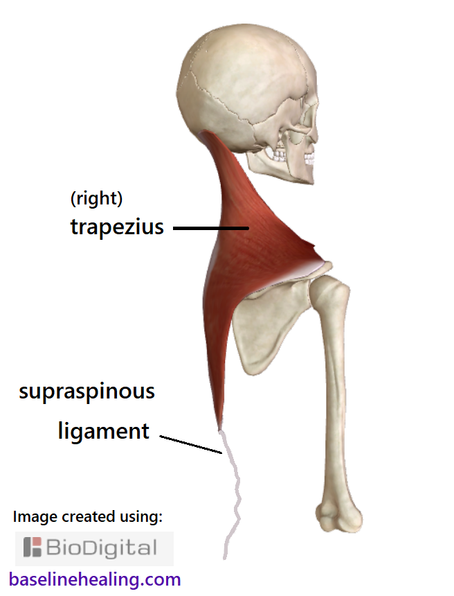
The left and right trapezius muscles emerge from the nuchal ligament and the thoracic portion of the supraspinous ligament. The nuchal ligament forms a septum - " a dividing wall" - between the upper parts of the trapezius muscles.

trapezius

By focusing on the condition and activation of the trapezius muscles, the positioning of the nuchal ligament can be appreciated.

off-center front view of the trapezius muscles and where they attach to the base of the skull and left and right scapula and collar bone. The cervical vertebrae have been removed so the nuchal ligament can be seen between the trapezius in the back of the neck.

You should be able to easily feel the nuchal ligament in your neck (I could not due to the restrictions in surrounding tissues.)

Extend your head backward and press your fingers on the midline of the back of your neck. Then tilt your head forward and should be able to feel the nuchal ligament 'popping out' as it tightens to limit the forward bending of your head and neck.

Supraspinous Ligament.

Supra ~ over/above + spinous ~ spine.

Continuous with the nuchal ligament, the supraspinous ligament is part of our midline anatomy. It is a strong, fibrous cord that attaches to the posterior spinal column, from the base of the neck to the lower back.

The supraspinous ligament attaches to each vertebra from the last cervical, all 12 thoracic vertebrae and the lumbar vertebrae to l3 l4 or l5.  The supraspinous ligament attaches to each spinous process that sticks out from each vertebrae.  Like a rope over a row of pillars on the median plane.

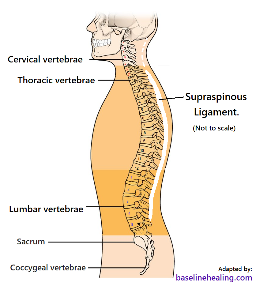
The supraspinous ligament attaches to the spinous processes of:

  • The seventh cervical vertebra.
  • The twelve thoracic vertebrae.
  • The upper lumbar vertebrae, usually terminating at L3, L4 or L5
  • .

The collagen fibres of the supraspinous ligament are arranged in bundles and layers. The deepest fibres connect to the spinous processes of adjacent vertebrae, the middle fibres run between 2 and 3 vertebrae and the most superficial fibres span 3 or 4 vertebrae.

At the points of attachment to the tips of the spinous processes fibrocartilage is developed in the supraspinous ligament and it is intimately blended with the interspinal ligaments and neighbouring fascia.

The trapezius muscles meet midline, merging with the supraspinous ligament from C7 to T12.

the trapezius muscles from neck to mid-back and the supraspinous ligament continuing to the lumbar area, a thin cord down the midline of the back of the spine.

The Nuchal & Supraspinous Ligaments

and Body Alignment.

The nuchal and supraspinous ligaments are the body's secondary anatomical guides for body alignment.

body alignment and balance

These midline structures should be free to fully extend and align with the linea alba (between the rectus abdominis muscles) on the median plane.

linea albathe median plane

Focusing on activating the trapezius muscles allows us to feel the relative positioning of the nuchal and supraspinous ligaments and work towards body alignment.

the nuchal ligament attaching to the back of the skull, running down the back of the neck, becoming the supraspinous ligament to the sacrum.  The left and right trapezius muscles attach to the nuchal ligament and thoracic part of the supraspinous ligaments on the median plane, our midline.  The nuchal and supraspinous ligaments should align with the linea alba at the front of the body between the rectus abdominis muscles. Note the size and shape of the trapezius muscles - large, kite-like sheets of muscle that extend out towards the shoulders from midline head to mid-back.

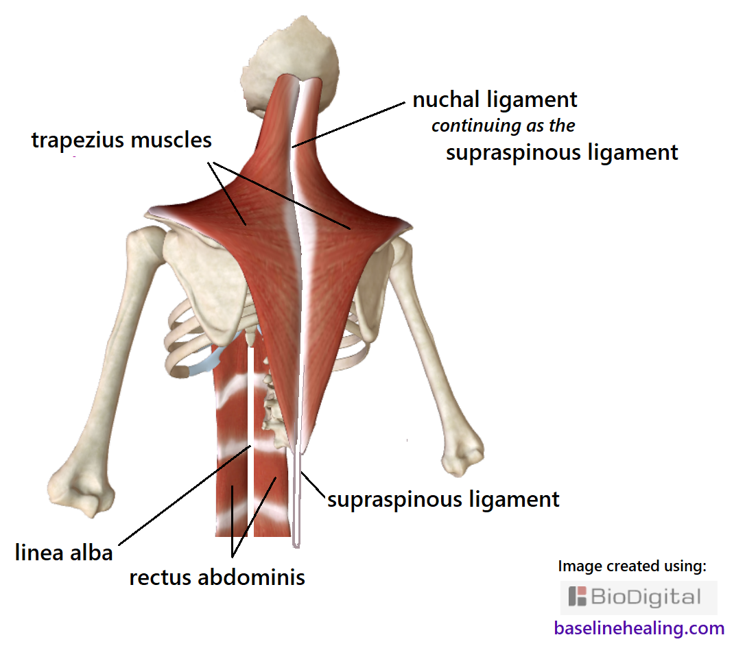
Alignment of the linea alba, nuchal and supraspinous ligaments is possible when the body is free of restrictions and has a full range of natural movement.

full range of natural movement

Back To Top